


Trans Vaginal Sonography
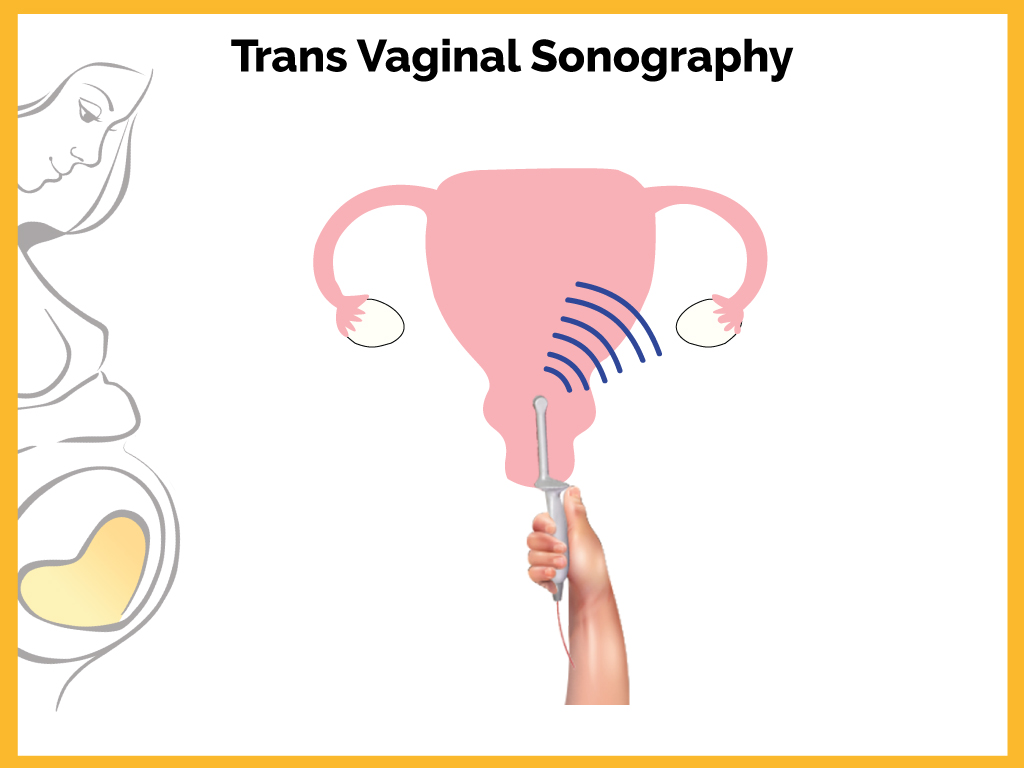
A transvaginal sonography or ultrasound, also called an endovaginal ultrasound, is a safe procedure used by doctors to examine internal organs in the female pelvic region. High-frequency sound waves create images of the uterus, fallopian tubes, ovaries, cervix, and vagina for the doctor to identify and diagnose conditions that hamper fertility. Unlike X-rays, ultrasound scanning techniques do not use radiation, which means that they have no harmful side effects and are very safe.
Who requires to undergo transvaginal ultrasound performed?
-
Transvaginal ultrasound is required if one
-
Needs an abnormal pelvic or abdominal exam
-
Has unexplained vaginal bleeding
-
Requires interventional procedures
-
Suffers from pelvic pain
-
Has early pregnancy
-
Is obese or gaseous
-
Has an ectopic pregnancy
-
Has pelvic masses
-
Suffers from infertility
-
Needs to undergo follicle monitoring checkup
-
Needs to undergo oocyte retrieval
-
Has retroverted or retroflexed uterus
-
Needs to undergo endometrial study to assess suitable IVF technique
-
Needs a check for cysts or uterine fibroids
-
Needs verification that an IUD is placed properly
The procedure
The patient is told to lie down on her back on the examination table and bend her knees. The doctor covers the ultrasound wand with a condom and lubricating gel, and then inserts it into the vagina. Between uses the probe should be soaked in disinfectant. If the patient has any latex allergy, a latex-free probe cover should be used. The patient might feel some pressure when the doctor inserts the transducer. The pressure is similar to the one felt during a pap smear when the doctor inserts a speculum into the vagina. Once the transducer is inside, sound waves bounce off the patient’s internal organs and transmit pictures of the interior of the pelvis onto a monitor. The technician or doctor then slowly turns the transducer, which provides a comprehensive picture of the organs.
The doctor might also suggest a saline infusion sonography, an ultrasound procedure that involves inserting sterile salt water into the uterus before the ultrasound to help identify any possible abnormalities inside the uterus. The saline solution stretches the uterus slightly, providing a more detailed picture of the inside of the uterus than a conventional ultrasound.
Preparation for transvaginal ultrasound
Depending on the doctor’s instructions and the reasons for the ultrasound, the patient’s bladder must be empty or partially filled. A full bladder helps lift the intestines and allows a clearer picture of the patient’s pelvic organs.